Eye Conditions
Corneal Sequestrum
Corneal sequestrum is a disease affecting the cornea (the clear curved transparent part of the front the eye). The sequestrum is usually an area of degenerated or non-living corneal tissue. This develops after an area of chronic corneal irritation or non-healing ulceration. The area of corneal degeneration often turns light brown to dark brown or black in color. The sequestrum may affect only the outer layers of the corneal stroma (tissue), but in some cases the sequestrum extends deeper into the cornea and may lead to deep ulceration, pain and possibly, corneal rupture.
While corneal sequestra have been documented in horses, the disease is most common in cats. Corneal sequestra occur in cats of all breeds, however, the Persian, Himalayan, and Burmese are particularly susceptible.
If your cat develops a corneal sequestrum, you might notice increased tearing, discharge from the eye and squinting. You might also see the brown (or black) area on the cornea.
A corneal sequestrum can lead to corneal ulcers (superficial to deep), ocular infections, corneal scarring, corneal vascularization and corneal mineralization.
Causes
Corneal sequestra can occur as the result of chronic eye irritation. A large prominent globe, low tear production, decreased blink response and decreased sensation of the cornea, as seen in brachycephalic (or short-nosed, flat-faced) breeds might create a predisposition to a corneal sequestrum. Another possible cause of this disease is Feline Herpesvirus. Sequestra can recur in the same eye or in the other eye in predisposed patients.
Treatment
Treatment for a corneal sequestrum usually involves a surgical procedure called a keratectomy. This involves surgical removal of the diseased layers of cornea. Our veterinary ophthalmologists perform this surgery using an operating microscope and specialized microsurgical instruments. Surgery relieves the discomfort associated with the corneal sequestrum, prevents the lesion from deepening and greatly shortens the healing time of the ulcer.
If the sequestrum affects the deeper layers of the cornea we may elect to apply a tissue graft or similar material to strengthen the cornea in that area and hasten healing. This may involve the use of a graft consisting of the patient’s own conjunctiva (the pink, mobile layer covering the white of the eye), adjacent cornea, donor cornea, synthetic biological or collagen discs placed over the surgical defect.
Not all corneal sequestra require surgical intervention. Your veterinary ophthalmologist may recommend medical treatment and monitoring. When surgical intervention is recommended, your doctor will discuss the surgical procedure along with benefits and risks specific to your pet. Any surgical procedure can introduce complications, including potential anesthetic risks. Surgical procedures that involve the cornea seldom give rise to complications, which occur in less than 5% of these cases. Nevertheless, potential complications include, but are not limited to:
•Inflammation of the pink tissue (conjunctivitis)
•Break down or rejection of the tissue or suture (wound dehiscence/graft retraction/rejection)
•Infections at the surgical site, which may extend to other internal and/or external areas of the eye
•Corneal ulcerations
•Corneal scarring, vascularization, or mineralization
•Inflammation inside the eye (uveitis)
Some of these complications, if severe could lead to blindness; however most can be managed with prompt treatment
If you have any further questions or concerns regarding corneal sequestrum, please do not hesitate to call us at Eye Care for Animals.
Less InfoEyelid Agenesis
General Information
Eyelid agenesis is a congenital condition (birth defect) in cats where a portion of the eyelid margin is underdeveloped at birth. This disease of the feline eyelid is also referred to as an “eyelid coloboma”, as a coloboma implies an area of missing tissue. This condition generally affects both eyes and most commonly occurs along the upper and outer aspects of the eyelids. The cause of eyelid agenesis is not fully understood; however, hereditary patterns or in utero viral transmission have been proposed. All cats with eyelid agenesis should receive a thorough ophthalmic examination as additional intraocular abnormalities may be present, which could lead to discomfort or vision impairment.
Treatment Strategies
Eyelid agenesis is generally accompanied by varying degrees of corneal irritation (keratitis) due to hairs rubbing on the cornea or improper tear film distribution leading to corneal drying. These side effects can occasionally lead to corneal ulceration varying from superficial to deep wounds.
- Medical Treatment –
- If the eyelid defect is small, and the cornea is healthy, medical treatment consisting of frequent application of ophthalmic lubricants may be sufficient. The goal of lubrication is to minimize the discomfort of both corneal drying (desiccation) and misdirected hairs, which may touch the corneal surface.
- Cryotherapy –
- The goal of cryotherapy is to freeze and remove misdirected hairs that are causing irritation or ulceration of the cornea. This procedure does not correct the eyelid malformation, but is designed to improve patient comfort, minimize secondary complications of eyelid agenesis, and reduce the amount of topical medications necessary.
- Reconstructive Eyelid Surgery –
- For larger defects, surgical reconstruction of the upper eyelid may be indicated for ocular health and comfort of your cat. There are several surgical methods available, and most are geared at relocating a normal piece of tissue from nearby structures (lower eyelid, lip, etc.) and grafting a “new” eyelid into the defect. The goal of this surgery is to recreate a new eyelid and restore normal function thus providing long-term comfort for your pet.
If a procedure is indicated for your pet, common postoperative care may include the use of short-term systemic antibiotics, anti-inflammatories, and pain medication as well as topical therapy which may include eye drops or eye ointment. An Elizabethan collar should be placed following eyelid surgery to prevent self-trauma. Complications of surgery are uncommon but include wound infection, suture dehiscence, or persistent ocular irritation.
If you have any further questions or concerns regarding eyelid agenesis, please do not hesitate to call us at Eye Care for Animals.
Less InfoFeline Conjunctivitis and Keratitis
Conjunctivitis and keratitis are common eye problems in all ages of cats. Conjunctivitis refers to the inflammation of the pink membranes surrounding the eye, whereas keratitis refers to the inflammation of the cornea. Cats may either develop signs of conjunctivitis alone, or symptoms coupled with keratitis, which is then termed keratoconjunctivitis. A number of infectious agents, namely viruses and bacteria, have been identified as potential underlying causes of these two conditions. These infections may be contracted as a kitten from the queen shortly after birth or from other cats, and are usually contagious from cat to cat.
Clinical Signs
The most common clinical signs of feline conjunctivitis and keratitis include red and swollen conjunctiva with excessive tearing, ocular discharge, and/or squinting. The discharge ranges from clear mucous to green- yellow in color. Some cats also rub at the eyes, which indicates discomfort. Sneezing and nasal discharge may also accompany the ocular signs, especially in younger cats. Recurrent bouts of conjunctivitis and keratitis are common, especially in viral cases such as feline herpes virus. Severe infections can cause complications such as permanent scarring of the cornea and conjunctiva. Some cases may have poor tear drainage resulting in chronic tearing onto the face. In young kittens, the inflammatory response may cause adherence of the conjunctiva to the cornea, and the eyelids may scar and fuse partially closed. The most severe cases can lead to rupture of the globe and vision loss, especially if untreated.
Severe keratitis can lead to abnormal blood vessel growth into the cornea. When severe infections occur, such as simultaneous viral and bacterial infections, corneal ulceration (a scratch or abrasion) may lead to perforation of the eye. With long-standing ulcers that are slow to heal, a dark brown to black discolored area of the cornea (corneal sequestrum) can form. When corneal ulceration and/or sequestrum occur, they are usually associated with varying degrees of ocular discomfort or pain.
Diagnosis
In addition to history and clinical signs, several diagnostic tests may be required to confirm the cause and magnitude of conjunctivitis and keratitis. When a cause is identified, specific treatment directed at the underlying cause produces a faster healing response. Some or all of the following tests may be necessary.
Conjunctival Cytology
Microscopic evaluation of the cells collected from the ocular surface can help determine the health of the cornea and conjunctiva. Furthermore, it will help identify whether primary or secondary infections are involved. For suspected viral infections, samples can be sent for virus isolation, fluorescent antibody (FA), and/or polymerase chain reaction (PCR) testing by a diagnostic laboratory. Although these tests are highly specific, they are not 100 percent sensitive; therefore, repeated samples may need to be submitted to the lab to achieve a positive result for a definitive diagnosis.
Cultures
Bacterial culture is often warranted to confirm suspected infections. In order to determine if the right antibiotic is used for a bacterial infection, the diagnostic laboratory can perform an antibiotic sensitivity test for the offending bacteria.
Blood Tests
Ocular disease can be the result of internal or systemic conditions; if the patient’s immune system is suppressed or not fully functional, infections are more likely to occur. Specific tests to check the health status of the patient may include a complete blood count, serum chemistry profile, tests for feline leukemia virus (FeLV), and feline immunodeficiency virus (FIV). Blood testing is also available for feline herpes virus.
Common Causes and Treatment
Common causes of conjunctivitis include feline herpesvirus (FHV-1), chlamydial, and other bacterial infections; a less common cause is allergies. Depending on the cause, certain antibiotic and antiviral agents may be used for treatment. Bacterial and chlamydial infections usually respond well to topical and oral medications. On the other hand, patients infected with viral agents may not respond as well to these treatments. Antiviral therapy requires frequent applications, sometimes as often as every 2-3 hours, in order to achieve the maximal effect. Antibiotic therapy is usually given at the same time as antiviral therapy in order to treat any secondary bacterial infection that may be present. Similar to human cold sores, viral infections are associated with stress; therefore, maintaining a low-stress environment may help reduce recurrences.
Superficial corneal ulcers can usually be treated similar conjunctival abnormalities; however, a large or deep corneal ulcer and corneal sequestrum may require surgical intervention. One specific treatment is surgical removal of infected or devitalized tissue (keratectomy). The cornea may require additional support, such as conjunctival grafting, following the keratectomy. Grafting also enhances the long-term health of a devitalized cornea by introducing blood vessels into a normally avascular area. With chronically infected eyes that contain excessive scarring, scar tissue may be surgically reduced to improve vision or tear drainage.
Prognosis
In general, response to therapy is good with bacterial and chlamydial conjunctival infections. Unfortunately, viral infections may be slow to heal and often recurrent. Because complications can have devastating consequences, such as pain and blindness, it is important that the treatment schedule is followed closely. Re-examinations to monitor progress and to adjust therapy will be advised when necessary.
If you have any further questions or concerns regarding feline conjunctivitis and keratitis, please do not hesitate to call us at Eye Care for Animals.
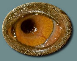
Less InfoFeline Diffuse Iris Melanoma (FDIM)
The iris is the most common origin of primary intraocular tumors in all species of domestic animals. The iris is the most common site of origin for ocular melanocytic (brown pigmented) tumors in canine and feline species. Feline diffuse iris melanoma is the most common type of ophthalmic melanoma (cancer) in cat species, with an average onset age of 11 years. FDIM begins as concentrated or multiple areas of iridal pigmentation that gradually increase in size.
The progression rate of FDIM is highly variable and the disease can have long dormant periods. FDIM can be metastatic (spreading of the tumor to the rest of the body) in felines and may spread to the lung, spleen, liver and other parts of the abdomen. This unpredictability makes it challenging for Veterinary Ophthalmologists to offer advice on when best to remove the affected eye, which may still provide vision.
Photographing the affected eye over time intervals is a very helpful way to document growth of the lesion, iridal pigmentation change, and changes in anterior chamber depth. Other diagnostic options include a full body X-ray to rule out the metastasis and a full blood panel to establish a baseline.
Laser therapy is not recommended for iris melanoma in cats owing to their higher metastatic potential.
Less Info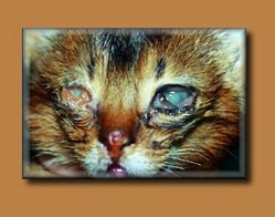
Feline Herpesvirus
Feline herpesvirus (FHV-1) is a common cause of eye and upper respiratory infection in the cat. This virus is very common in the cat population, but it is not contagious to people and other species of animals such as dogs. Herpesvirus is easily passed from one cat to another through sneezing, coughing, grooming, and/or simply living in the same household with an infected cat. While it is quite contagious, most cats contract this infection from their mothers before they are even weaned. This virus is in the same family as the chicken pox virus. As you may know, some people will develop ‘shingles’ in their adult lives, which is a re-emergence of the chicken pox virus that has been lying dormant in the body since childhood. The same is true for cats with FHV-1. Clinical signs associated with infection can vary greatly between cats. Some cats will never have signs after the initial infection while other cats will have episodes throughout life.
Some cats affected with FHV-1 may only have mild conjunctivitis (redness and inflammation of the white part of the eye) of one or both eyes. Other cats have more severe disease and may show ocular and nasal discharge, conjunctivitis, coughing and sneezing. Cats may also develop ulcers of the cornea (the clear “window” in the front part of the eye). Corneal ulcers can be very painful and serious enough to cause noticeable scarring or even perforation of the normally clear cornea.
After initial recovery from the herpesvirus infection, an estimated 80% of the cats become a carrier of the disease. In other words, the disease goes into temporary remission but the cat may still be infectious. In these cats, stress and illness can reactivate the virus, causing repeated infections or recurrences of the clinical signs throughout life. Stressors may include moving to a new house, having new pets or houseguests in the household, construction/remodeling, a traveling owner, etc.) Repeated or chronic infections have been associated with diseases such as dry eye, symblepharon (adhesion of the conjunctiva to itself or to the cornea), corneal sequestrum (an abnormal brown plaque formation on the cornea), and possibly eosinophilic keratitis (an immune-mediated condition of the cornea).
The definitive diagnosis of feline herpesvirus infection is accomplished by laboratory testing. Many tests are available through a professional diagnostic laboratory. They include virus isolation, fluorescent antibody (FA) testing, serology such as ELISA or serum neutralizing titers, and polymerase chain reaction (PCR) testing. It is important to realize that these tests may have false negative results. On the other hand, certain cats in the carrier state but without apparent clinical signs can also test positive for some of these diagnostic modalities. Since the disease is extremely common, it is not always necessary to perform diagnostic testing, but your veterinary ophthalmologist will discuss this with you at the examination.
Treatment for herpesvirus is aimed at controlling clinical signs and reducing secondary complications. It is important to note that there is no cure for herpesvirus, and once infected, your cat has the virus for life. Some animals will never have clinical disease after the initial infection while others may have frequent recurrences. Cats that have recurrent outbreaks often have a stressful trigger, which if identified can be avoided or minimized. This can reduce the number of outbreaks. Typically, therapy includes topical antiviral drops or ointment for the eye and occasionally an oral antiviral medication. Sometimes starting medications prophylactically (before a known stressor) the severity of the recurrent infection can be reduced. Some anecdotal reports state that L-lysine, an amino acid dietary supplement, can inhibit viral replication. This has been shown in a laboratory setting but not in cats with natural infection. There are no studies proving that Giving L-lysine as a supplement may benefit cats with herpesvirus as many owners feel it reduces outbreaks. In any event, there are no known side effects of L-lysine documented in the cat.
Vaccination against herpesvirus infection is included in the typical feline vaccination schedule provided by your primary care veterinarian. The vaccine minimizes the clinical signs of the herpesvirus infection but does not prevent future outbreaks. Additionally, the vaccination does not cure cats already infected with the herpesvirus.
If you have any questions or concerns regarding feline herpesvirus, please call us at Eye Care for Animals.
Less Info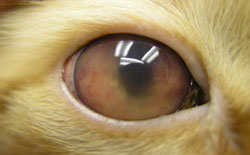
Feline Uveitis
Uveitis is an inflammatory process involving the middle of the three layers in the eye. To understand uveitis, it is important to know the basic anatomy of the eye. The outer layer enclosing the eye is composed of the clear cornea and the white sclera. The innermost layer is the nerve layer or the retina. The middle layer is the uveal tract, which is rich in blood vessels. It is composed of the iris in the front part of the eye, the ciliary body, which produces the fluid (aqueous humor) inside the eye, and the choroid which nourishes the retina in the back of the eye. Because of its rich blood supply, the uveal tract is a natural target for diseases originating in other parts of the body. When inflammation attacks specific segments of the uveal tract, the disease is further classified as iritis (inflammation of the iris), cyclitis (inflammation of the ciliary body) or choroiditis (inflammation of the choroid), depending on the affected structure. If all the structures are inflamed then it is called panuveitis (inflammation of all uveal structures of the eye).
Diagnosis of Uveitis
Ocular pressure is maintained by the aqueous humor (fluid) produced by the ciliary body within the eye. Initially, if the ciliary body is inflamed, the fluid production slows down and the ocular pressure drops. The aqueous humor produced in the eye normally drains through the angle between the ciliary body and the iris. The inflammatory debris produced in uveitis can block the drainage angle and result in increased intraocular pressure (glaucoma) over time. Once uveitis resolves, glaucoma can remain if drainage structures were damaged by the inflammation. A recheck of the eyes following uveitis is important for this reason.
Additionally, disease processes such as uveitis can lead to lead to corneal ulcers (superficial to deep), ocular infections, corneal scarring, corneal vascularization, corneal mineralization, cataract, lens luxation, retinal detachment and keratoconjunctivitis sicca. Uveitis also can lead to secondary complications similar to those to which treatment for uveitis can give rise, as discussed under “Prognosis”.
Causes of Uveitis
Uveitis is associated with many different diseases. Examples in the dog include Ehrlichiosis and Coccidioidomycosis, two systemic diseases common to the southwestern United States. In the cat, uveitis can be a consequence of Feline Leukemia Virus, Feline Infectious Peritonitis or many other diseases. In any animal, penetrating injuries such as cactus spines or a cat scratch may produce uveitis. Inflammation of the uveal tract can occur when the lens capsule is breached (such as following surgery, trauma, or injury of the lens) or in the presence of cataracts where lens proteins leak out of the lens capsule into the eye. Other possible causes of uveitis are local bacterial infection, immune-mediated diseases and parasitic diseases. Treatment can be more specific if the actual cause is known. It is important to test for some infectious diseases to make sure there is not an underlying cause for the inflammation, but unfortunately, in up to 75% of the cases the cause is never determined.
Treatment
Uveitis must be treated aggressively in order to prevent glaucoma, scarring of the uveal structures, and possibly blindness. Different medications may be used to treat the underlying, original cause of the uveitis and to attempt to control the inflammation itself. Aspirin (not aspirin substitutes) and corticosteroids minimize the inflammatory process. Corticosteroids may be administered by injection under the lid of the eye, by drops in the eye, or as an oral medication, depending on the suspected cause of uveitis. Topical use must be postponed if damage to the corneal surface is present because the corticosteroids prevent healing of the ulcer. If certain systemic diseases are suspected, oral corticosteroids may be postponed until test results become available. Atropine dilates the pupil and helps prevent scarring of the iris. This medication may be contraindicated, however, if glaucoma is present as it may further decrease the drainage of aqueous humor from the eye. Oral and topical antibiotics are employed when a bacterial infection is present in the eye.
Prognosis
The treatment of uveitis requires therapy to halt the inflammation of the uveal tract along with a search for the original cause of the disease. Many tests may be needed to determine possible causes and the results are important for proper treatment.
Treatment for uveitis can involve life-long topical and/or oral medications. Life-long topical medications seldom give rise to complications, which occur in less than 5% of these cases. Nevertheless, potential complications include, but are not limited to, inflammation of the pink tissue (conjunctivitis); corneal ulcerations (superficial to deep); corneal scarring, vascularization, and mineralization; ocular rupture, secondary to ulceration; worsened inflammation inside the eye, secondary to infection or ulceration; high pressure within the eye (glaucoma), secondary to the uveitis; retinal detachment or degeneration, secondary to uveitis or infection; ocular or orbital pain, secondary to uveitis, glaucoma or infection; eyelid rubbing; bleeding inside the eye (hyphema), secondary to uveitis or infection; tearing (epiphora); and/or lens luxation, secondary to uveitis, glaucoma or infection. Some of these complications can lead to blindness. Some oral medications used to treat these disease processes may cause changes in behavior, gastrointestinal upset (diarrhea, vomiting, decrease/increase in appetite/thirst), panting, decreased white blood cell counts (such as leukopenia), and various changes in chemistry values (liver, kidney, potassium, to name a few).
Your awareness of your pet’s symptoms and compliance with recommendations for recheck examinations and periodic blood work help control these potential complications.
Less InfoFind a location
New Patients
Prepare for Your Visit:
New Patient FormEquine Client Info Form
Browse Eye Conditions
Referring Doctors
Educational MaterialsVet Referral Form